
Regular eye exams are vital to maintaining your vision and overall health and can help with the early and sight-saving diagnosis of critical eye conditions.
Many eye problems can develop without you knowing. You may not even notice any change in your sight. Fortunately, we can detect diseases or damage such as macular degeneration, glaucoma, retinal tears or detachments, and other health problems such as diabetes and high blood pressure with a thorough retina examination.
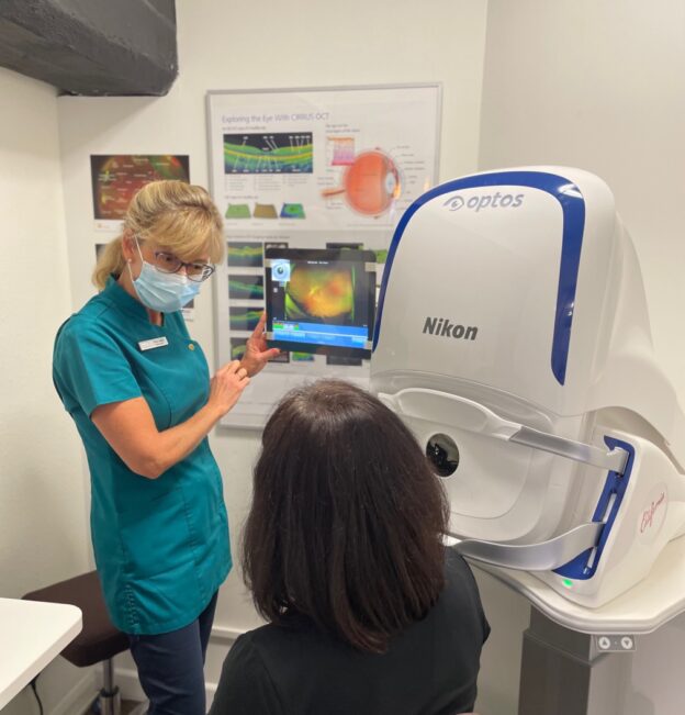
Here at Walker and Campbell, we offer the optomap as an essential part of our eye examinations. It provides us with a high-resolution 200° image, giving us a comprehensive view of your retina. Using optomap, we’ve been able to diagnose some critical eye conditions and help save our patients’ eyesight.
Four recent examples of critical conditions revealed using Optomap®
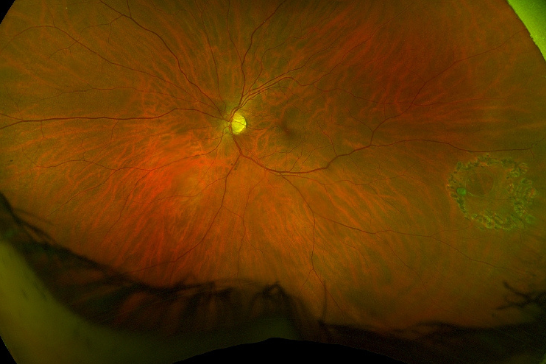
Retinal Detachment
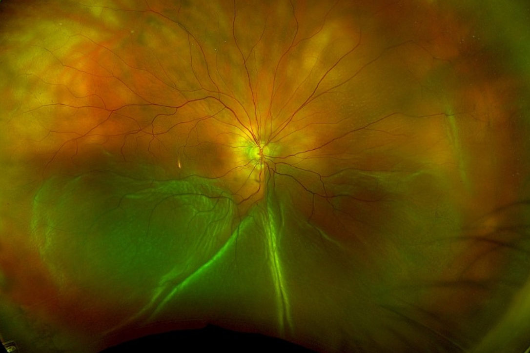
Retinal detachment
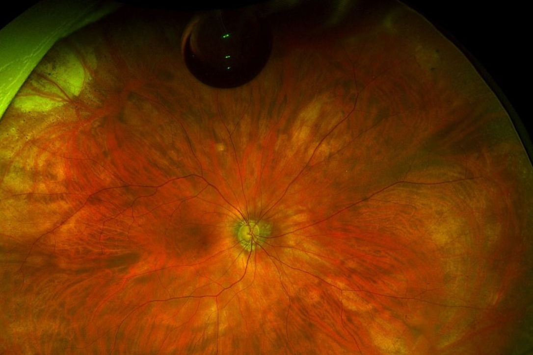
Retinal detachment post surgery
This first image is from a lady who came in because she thought she had a twig in her eye. An examination using Optomap showed the bottom half of her retina detached and flapping around, which could have caused total blindness if not treated. The second picture shows her retina nicely attached again with the gas bubble floating at the top.
Retinal Tear
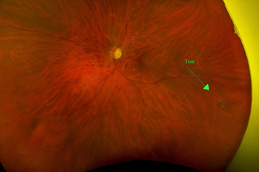
Retinal Tear
Retinal tear after laser surgery
The first picture shows a horseshoe-shaped tear in the periphery on the right-hand side which would certainly have developed into a detachment if not treated. The second picture shows laser burns around the tear to seal it down and stop it from progressing.
Diabetic Retinopathy
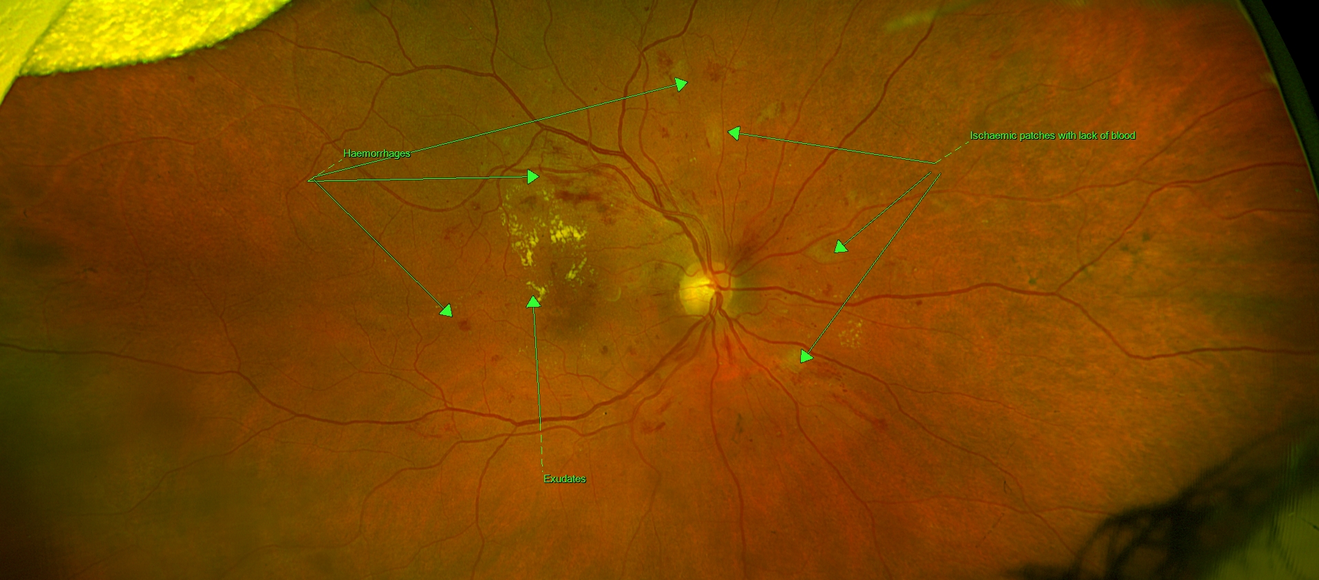
Diabetic retinopathy
This picture shows widespread haemorrhages. The bright yellow patches are exudates (fluid leaking from the blood vessels). The faint yellow patches are what we call ‘Cotton Wool Spots’ – these are ischaemic areas where the retina does not have a good blood supply which can cause tissue damage.
This patient was referred to their local Eye Hospital and is about to start having Eyelea injections to reduce the oedema (fluid leaking underneath the macula (central area of the retina).
Papilloedema
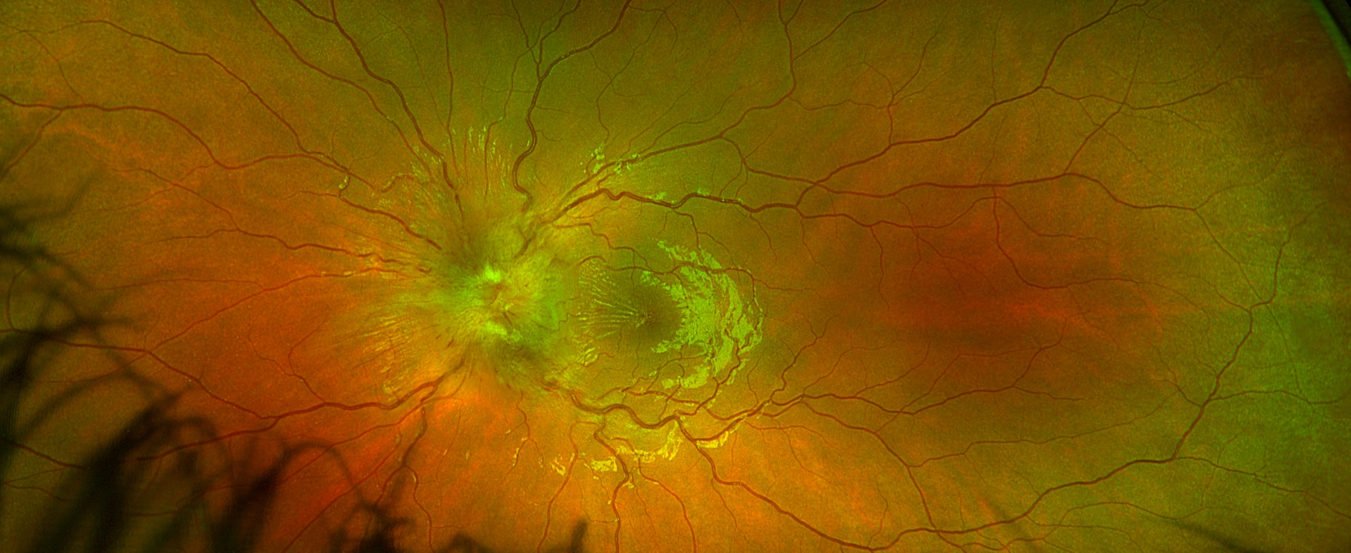
Papilloedema
This image shows papilloedema discovered during a routine eye examination of a 14-year-old patient.
Papilledema is a severe condition in which the optic nerve at the back of the eye becomes swollen due to a build-up of pressure in or around the brain. Often, it’s a warning sign of a critical medical condition that needs prompt attention, such as a brain tumour or haemorrhage.
We referred her to hospital, where she had many tests, including a CT scan and a lumbar puncture which confirmed that the problem was caused by raised intracranial pressure; luckily no tumour. She stayed in hospital for five days and was given medication to lower the pressure. No surgery was needed. She is now fit and well but must continue taking this medication for the foreseeable future. The family are all extremely grateful for our prompt detection and referral!
If you are concerned about your eyesight, have a family history of eye conditions, or haven’t had an eye examination in over two years, contact us to arrange an appointment for our comprehensive, advanced eye examination. Early detection could help save your sight.