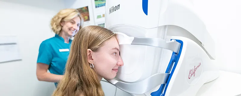
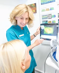
 Regular eye exams are vital to maintaining your vision and overall health. Walker and Campbell offers the optomap® as an important part of our eye examinations. The optomap produces an image that is unique and provides us with a high-resolution 200° image in order to ascertain the health of your retina. This is much wider than a traditional 45° image.
Many eye problems can develop without you knowing. In fact, you may not even notice any change in your sight. Fortunately, diseases or damage such as macular degeneration, glaucoma, retinal tears or detachments, and other health problems such as diabetes and high blood pressure can be seen with a thorough exam of the retina.
The inclusion of optomap® as part of our comprehensive eye exam provides:
- An image to show a healthy eye or detect disease.
- A view of the retina, giving us a more comprehensive view than we can get by other means.
- The opportunity for you to view and discuss the optomap® image of your eye with us at the time of your exam.
- A permanent record for your file, which allows us to view your images to look for any changes over time.
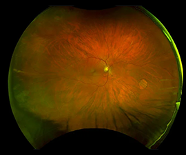
Glaucoma
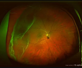
Retinal Tear
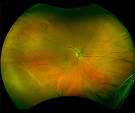
Diabetes
optomap® is fast, easy, comfortable and suitable for both adults and children. No dilation drops are needed. The entire imaging process takes place in our pre-test area and simply consists of you looking into the device one eye at a time. The images are shown immediately on a computer screen so we can review it with you.
Find out more about optomap® here.

